Epidermal Necrolysis
Severe immune-mediated epidermal necrosis and desquamation at ⩾2 distinct sites that is divided by extent of skin involvement into:
- Stevens-Johnson Syndrome
<10% BSA. - SJS/TEN Overlap syndrome
10-30% BSA. - Toxic Epidermal Necrolysis
>30% BSA.
Epidemiology and Risk Factors
Epidemiology:
- Annual incidence ~5/1,000,000.
Risk factors:
- ↑ Age
- Female sex
- HIV
- Connective tissue disease
- Malignancy
Pathophysiology
T-cell mediated reaction:
- Drug-specific CD8+ T-cells are activated
- Cytotoxic proteins released
- Epidermal necrolysis released
Aetiology
Precipitants include:
- Medications
Most cases are due to an idiosyncratic reaction to certain families of medications that:- Are usually commenced between 1 week and 1 month prior
- Include:
- Antibiotics
- Amoxacillin/ampicillin
- Fluoroquinolones
- Sulfamethoxazole
- Doxycycline
- Rifampicin
- Antiepileptics
- Carbamazepine
- Lamotrigine
- Phenobarbital
- Phenytoin
- NSAIDs
- Antibiotics
- Infection
- Mycoplasma
- Idiopathic
Assessment
- Identify the potential causative agents
- Assess the degree of skin loss
Assessment principles are similar to Burns.
History
Prodromal symptoms:
- Malaise
- Fever
- Sore throat
- Conjunctivitis
Examination
Mucocutaneous features:
- Lesions typically begin on the face and thorax
- Symmetrically distributed
- Typically begin as flaccid bullae
- Progress to “sheet-like” detachment of skin
- Mucosal involvement in 80%
- Genital involvement in 70%
- Corneal involvement in >60%
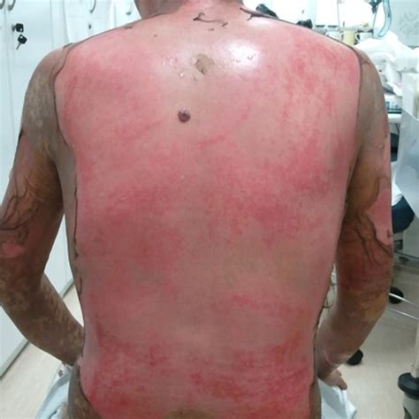
The Nikolsky sign is positive if epidermal detachment can be extended with gentle lateral pressure.
Investigations
Bedside:
Laboratory:
Imaging:
Other:
- Skin biopsy
Diagnostic Approach and DDx
Disease occurs in two phases:
- Acute phase
5-7 period of worsening desquamation and mucositis. Risk of:- Hypovolaemia
- Haemoconcentration
- AKI
- Sepsis
- Multiorgan failure
- Death
- Hypovolaemia
- Chronic phase
Convalescence and recovery.
Key differentials:
- Erythema multiforme
- Drug eruptions
Management
- Treat precipitant
- Transfer to burns centre
Resuscitation and supportive care has many similarities to burns, and is covered under Burns.
Resuscitation:
- A
- Intubation
May be required if mucosal involvement
- Intubation
Specific therapy:
- Pharmacological
- Cease offending agent
- Procedural
- Physical
- Specific ocular care
- Ocular rinses
- Lubricants
- Conjunctival adhesion separation
- Glass rod
- Forceps
- Specific genital care
Risk of fibrosis with adjacent skin edges, consider:- Topical corticosteroid
- Silicone vaginal dilators
- IDC placement
- Regular foreskin retraction
- Specific ocular care
Supportive care:
- D
- Multimodal analgesia
- F
- Volume resuscitation
Prevent hypovolaemia.
- Volume resuscitation
- G
- Early EN
May need 25-30kcal/kg/day due to ↑ metabolism. - Stress ulcer prophylaxis
- Early EN
- H
- Thromboprophylaxis
- I
- Wound dressings
- Non-adhesive
- Wound dressings
Disposition:
Preventative:
Marginal and Ineffective Therapies
Systemic immunomodulation therapy has limited evidence, however there is potential benefit from may:
- Pulse steroids
May be more effective when commenced early in disease onset. - IVIG
1-2g/kg/day. - Ertanercept
TNF-α inhibition.
Non-recommended agents include:
- Thalidomide
Anaesthetic Considerations
Complications
- Death
~25%, with ↑ mortality with ↑ desquamation. - B
- Lung injury
Direct bronchial sloughing. - HAP/VAP
- Lung injury
- F
- AKI
- Pre-renal
- AKI
- G