Chest
| Component | Inspection | Palpation | Percussion | Auscultation |
|---|---|---|---|---|
| General |
|
|||
| Respiratory |
|
|
|
|
| Cardiovascular |
|
|
|
|
| Neurological | ||||
| Endocrine and Metabolic | ||||
| Renal | ||||
| Gastrointestinal |
|
|||
| Haematological | ||||
| Integumentary | ||||
| Trauma |
|
|||
| Infective | ||||
| Malignant | ||||
| Toxic | ||||
| Immune | ||||
| Congenital | ||||
| Obstetric |
Features
Respiratory
- Chest form
- Barrel chest
Suggests hyperexpansion from COPD. - Scoliosis/kyphoscoliosis
Restrictive lung disease due to ↓ chest volume and ↓ chest wall compliance. - Funnel chest
Restrictive lung disease.
- Barrel chest
- Subcutaneous emphysema
Subcutaneous air, palpable in large volumes. Causes include:- Pneumothoraces
- Bronchial injury
- Hollow viscus injury
Rarely.
| Sound | Location | Timing | Nature |
|---|---|---|---|
| Tracheal breathing | Sternum/manubrium |
|
|
| Bronchial breathing | Parasternal |
|
|
| Vesicular | Peripheries |
|
|
Bronchial breathing may be physiological or pathological, depending on the location.
| Sound | Location | Timing | Nature | Aetiology |
|---|---|---|---|---|
| Bronchial breathing | Peripheries |
|
|
Consolidation of alveoli small airways between the chest wall and patent airways. Occurs as normal breath sounds are transmitted more readily through consolidated lung. |
| Absent | Anywhere |
|
|
↓ Airflow to affected side, which may indicate:
|
| Crackles | Peripheries |
|
|
Popping open of collapsed airways in early inspiration:
|
|
|
|
||
|
|
|||
| Ronchi | Peripheries |
|
|
|
| Wheeze |
|
Two forms:
|
||
| Pleural rub | Anywhere |
|
|
|
- Resonance
The sound produced by chest wall percussion can be divided into:- Normal
- Hyper-resonant
Higher-pitched, “hollow” sound, indicating air (classically bullae or pneumothoraces). - Dull
Lower-pitched, shorter sound, indicating effusion.
Percussion is performed with a finger of the non-dominant hand on the chest wall, and firmly tapping the distal segment with a finger on the other hand.
Remember to remove the percussing finder from the struck finder immediately - leaving the two fingers in contact ↓ the pitch of the produced note.
Cardiovascular
- Apex Beat
Palpation of the apical impulse, generated by the heart pushing into the chest wall during isovolumetric contraction.- The normal apex beat is:
- Brief
- “Tapping”
- Felt over a small area
- With LV dilatation, the apex beat:
- Moves laterally and caudally
- Can be felt over a larger area of the heart
- Becomes “heaving”
- With LV hypertrophy:
- The location is unchanged
- Becomes stronger
Described as “thrusting”.
- With a hyperdynamic circulation:
- May be seen as a sternal heave
- The normal apex beat is:
| Sound | Timing | Aetiology | Considerations |
|---|---|---|---|
| S1 |
|
Closure of mitral and tricuspid valves |
|
| S2 |
|
Closure of aortic and pulmonary valves |
| Sound | Timing | Aetiology | Considerations |
|---|---|---|---|
| S3 |
|
Rapid deceleration in diastolic filling due to diastolic dysfunction:
|
|
| S4 |
|
Vigorous atrial contraction |
Murmurs are additional heart sounds that occur due to pathologically turbulent blood flow.
| Timing | Lesions |
|---|---|
| Pansystolic |
|
| Ejection and midsystolic |
|
| Late systolic |
|
| Mid-late diastolic |
|
| Continuous |
|
- Cardiac wheeze
Bibasal expiratory wheeze secondary to ↑ extravascular lung water in dependent regions, leading to small airway compression.
Gastrointestinal
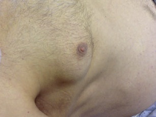
- Gynaecomastia
↓ Oestrogen clearance in men with chronic liver disease.
Trauma
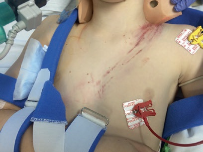
- Seatbelt sign
Chest (and/or abdominal) wall bruising associated with a 3-point restraint. Indicates a high-energy mechanism and a high risk of internal injury.
References
- Foot C, Steel L, Vidhani K, Lister B, MacPartlin M, Blackwell N. Examination Intensive Care Medicine. Elsevier Australia; 2011. (Examination series).